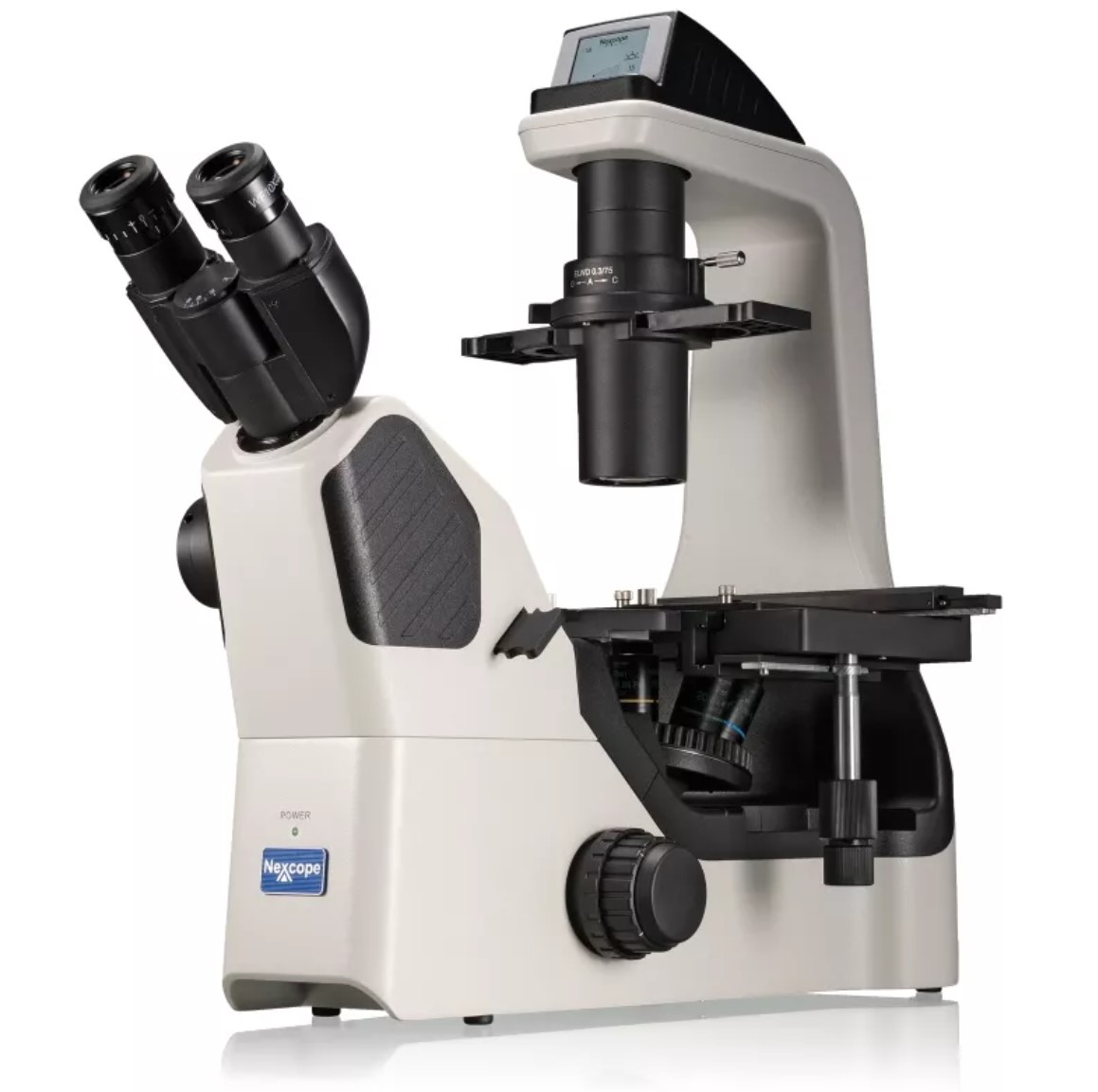
INVERTED BIOLOGICAL MICROSCOPE
NEXCOPE NIB600 Advanced Scientific Research Microscope for Cellular Observation
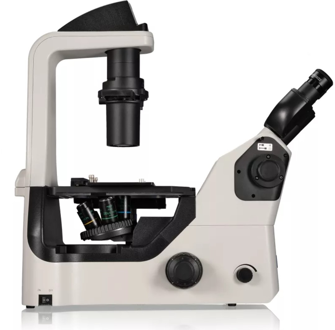
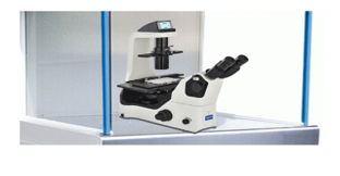
- The Nexcope NIB600 inverted microscope stands out with its sturdy body and base made of corrosion-resistant aluminum alloy. Its white epoxy electrostatic paint coating ensures durability. The microscope features anti-fungal treated optical sets for long-lasting use, and the NIS-60 objective set delivers superior image quality.
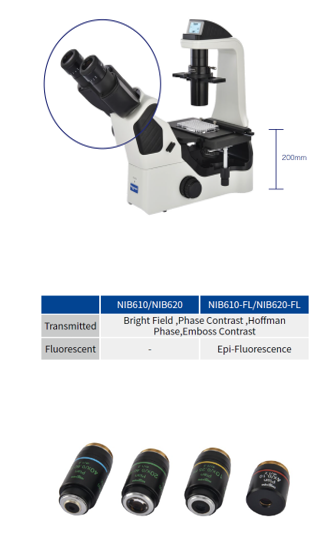
- The main body is designed with a modern and ergonomic structure, with an inverted design. For user comfort, it is equipped with a "Siedentopf" type binocular tube inclined at 45 degrees and an interpupillary distance adjustable between 48 and 75 mm. The tube also offers a side auxiliary port for connecting digital cameras and video recorders. Additionally, the image splitter system allows the optical path to be split at a ratio of 100/0 or 0/100.
- The optical system includes 10X eyepieces with a wide field of view, and these eyepieces can be adjusted individually for each eye using diopter control (+5/-5 degrees). The eyepieces are also equipped with reticle holders and indicator needle protection.
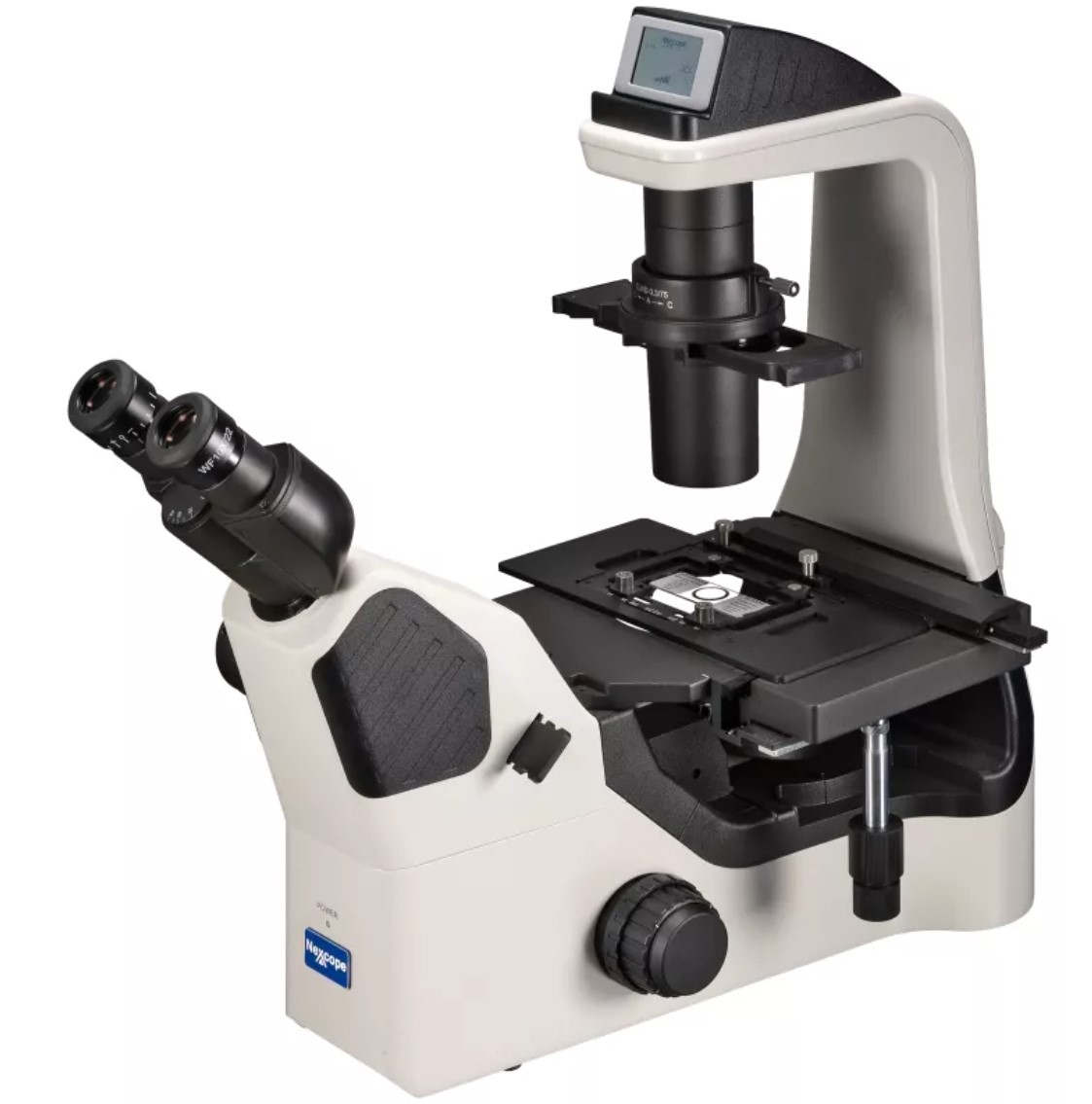
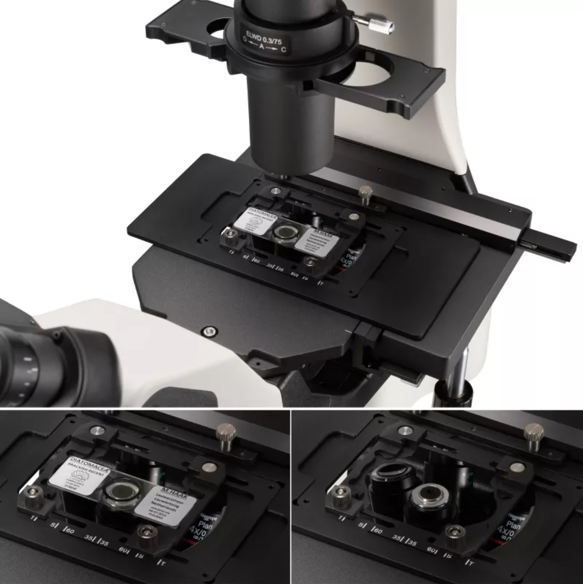
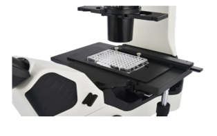
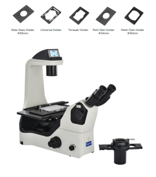
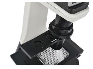
- The microscope has a fixed rectangular stage measuring 250 mm x 170 mm, along with an integrated Vernier scale. The ergonomic Charriot control system on the X and Y axes provides precise movement. The universal F/TE sample clamp allows for secure placement of various sample types; Petri dishes, hemocytometers, and other sample containers of different sizes can be easily accommodated.
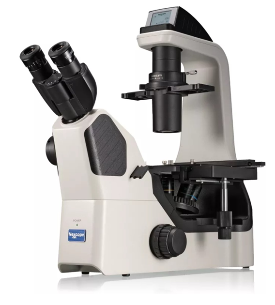
PROFESSIONAL CELL IMAGING
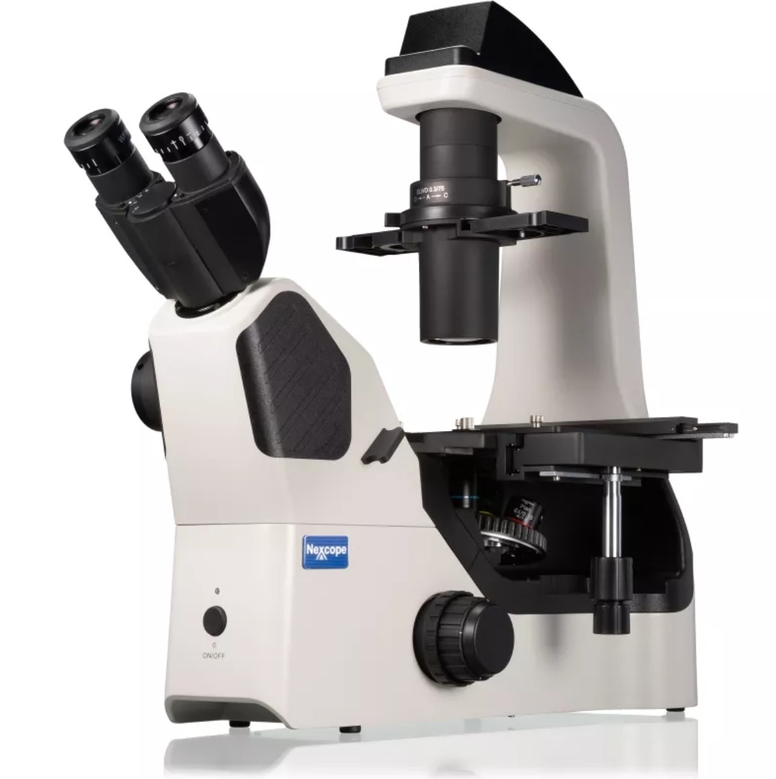
Ergonomic Design, Comfortable Use
45° Inclined Viewing Head
The inclined viewing head allows the user to operate the microscope in a comfortable position. It minimizes muscle tension and discomfort caused by long working hours.
Long-Handle Mechanical Stage
The user can make smooth and comfortable movements during the operation, thereby improving work efficiency and comfort.
High Brightness, Long-Life LED Illumination
The high-brightness and long-life LED illumination system provides uniform brightness and cool light for both transmitted and fluorescent lighting.
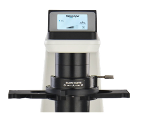
Objective Coding Converter
It can memorize the illumination brightness when each objective is used. When different objectives are switched, the light intensity is automatically adjusted to reduce visual fatigue and improve work efficiency.
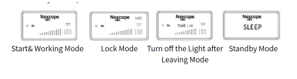
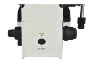
Use the Adjustment Knob to Perform Multiple
FunctionsClick: Enter standby mode
Double click: Lock or unlock the light
Rotate: Adjust brightness
Up-spin: Switch to the upper light source
Down-spin: Switch to the lower light source
Press for 3 seconds: Set the light off time
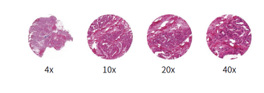
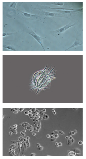
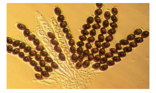
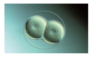
PHASE CONTRAST
By utilizing changes in the refractive index, high-contrast microscopic images of transparent samples can be obtained using the phase-contrast observation technique. The advantage is that live cell images can be obtained without the use of staining or fluorescent dyes.
Application areas:
- Living cells in culture
- MicroorganismsDoku preparatları
- Subcellular structures (including cell nuclei and organelles)
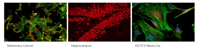
FLUORESCENCE OBSERVATION
LED lighting makes fluorescence observation easier.
Uniform Brightness
In accordance with Kohler illumination, the Fly-eye lens ensures uniform brightness across the entire field of view when observed through the eyepieces or CCD camera.
Ease of Use
Compared to traditional mercury lamps, LED lighting eliminates the need for frequent bulb replacements, saving time and money. It also resolves issues related to heating, cooling, and high temperatures.

It is suitable for many different fluorescent dyes.
It is equipped with 3 fluorescent filter blocks, providing a wide range of dyes. This ensures clear and high-contrast fluorescent images.
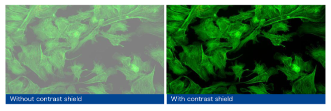
CONTRAST PROTECTION
Contrast protection effectively prevents interference from external light, enhances the contrast of the fluorescent image, and provides fluorescent images with a high signal-to-noise ratio. When phase contrast observation is required, the contrast protection can be easily removed from the light path, preventing any negative effects on the quality of the phase contrast.