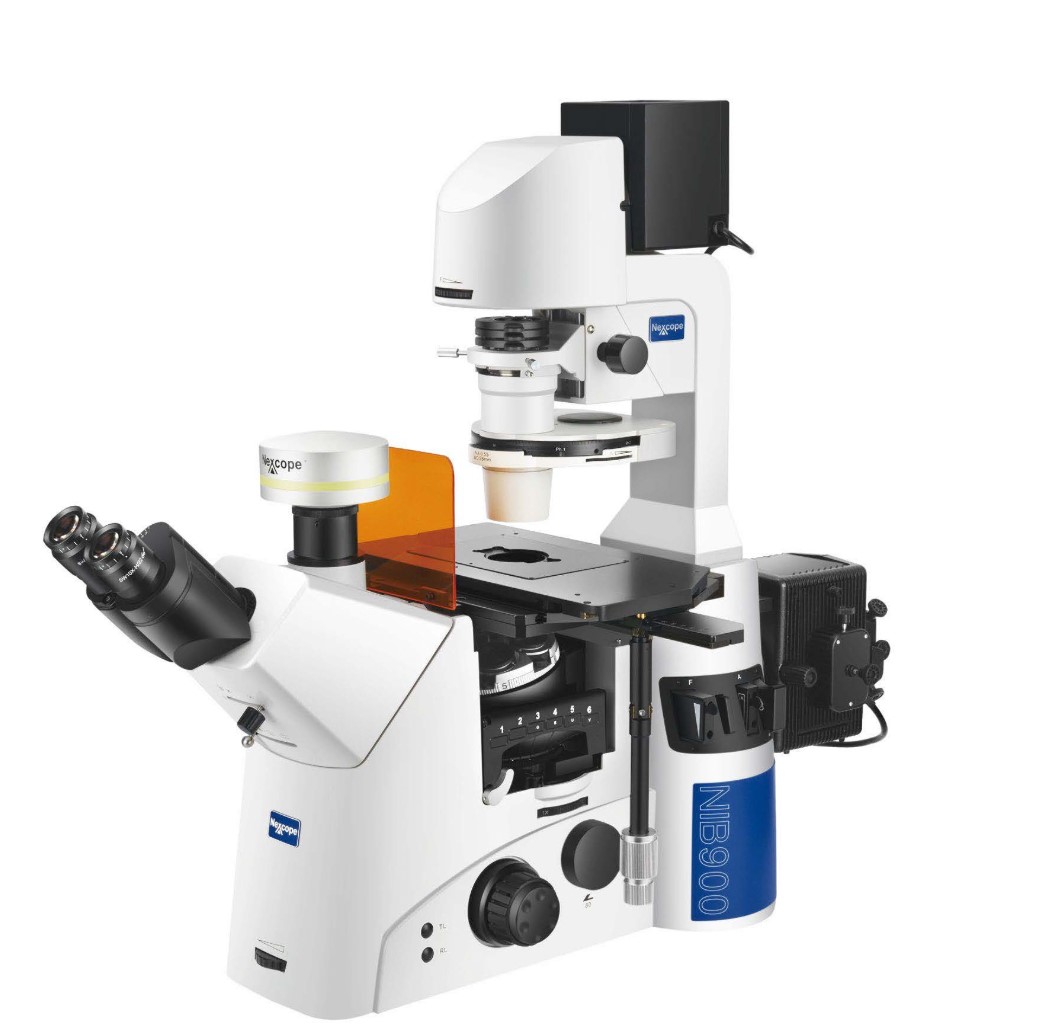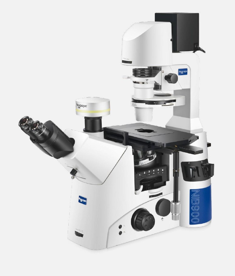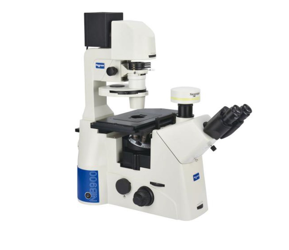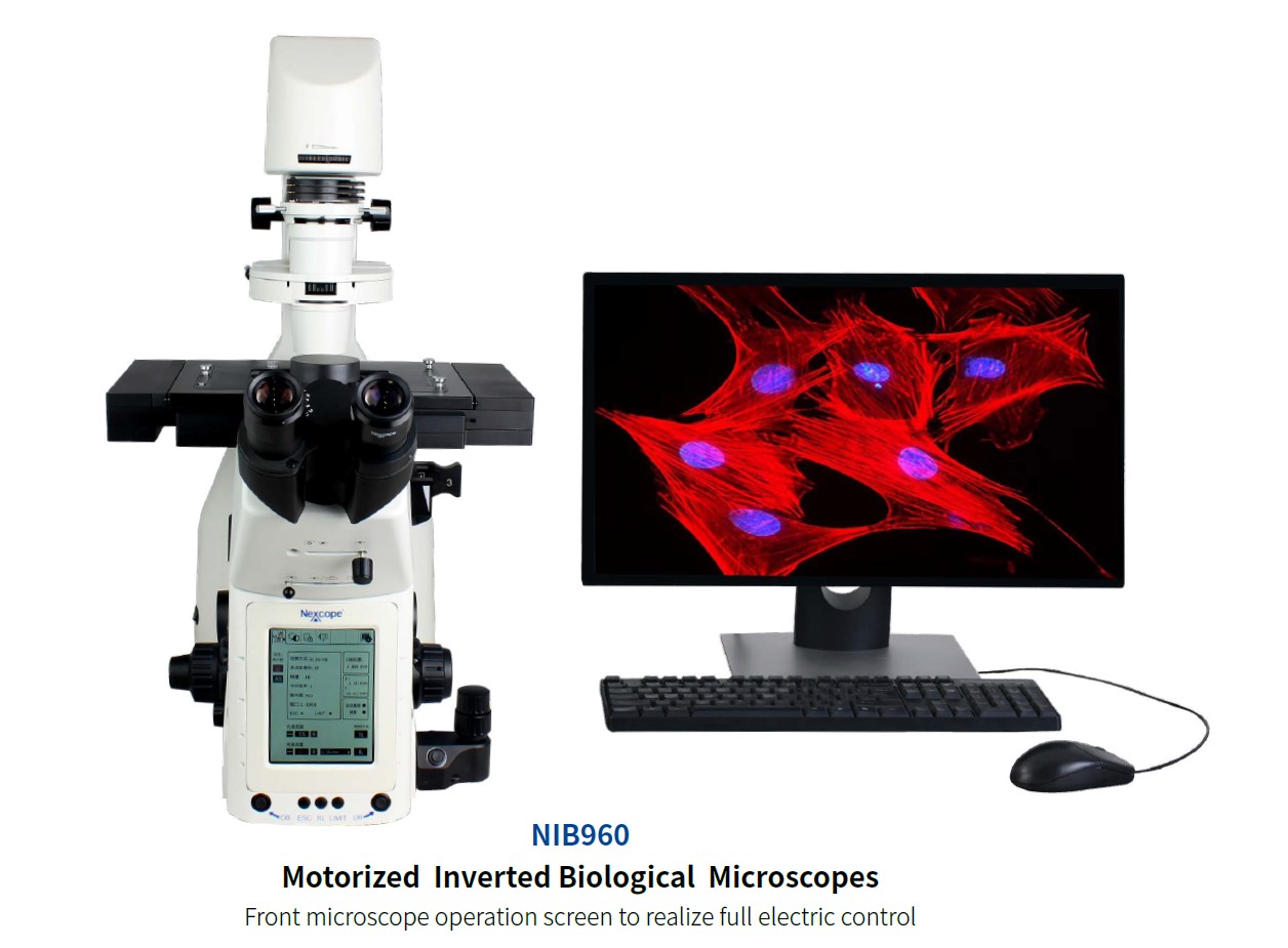
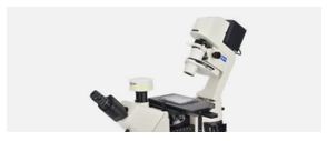
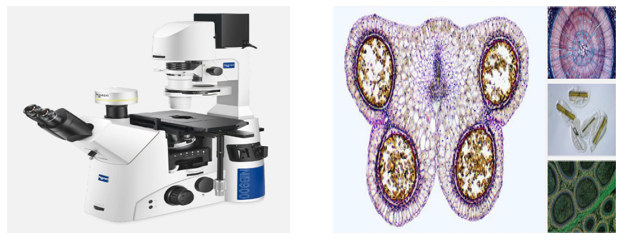
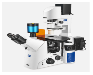
INVERTED RESEARCH MICROSCOPE
NEXCOPE NIB900 Brightfield, Darkfield, Phase Contrast, DIC, Fluorescence, Polarized
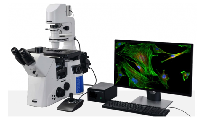
- The NIB900 series, an inverted microscope designed to meet the demands of advanced life science research, can address a wide range of needs. This versatile microscope is capable of performing various observation methods such as brightfield, phase contrast, polarization, DIC, fluorescence, and more. It also supports confocal and super-resolution imaging, which is essential for cutting-edge life science research.
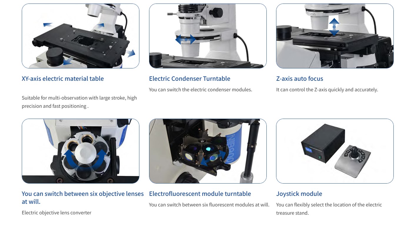
- The ergonomic design of the NIB900 series significantly reduces mechanical fatigue caused by long observation sessions by fully considering the user's work habits. Additionally, by adopting high-speed motorized control to simplify and facilitate complex operations, it makes the overall workflow easier and more intuitive.
- This combination of versatility, precision, and user-focused design makes the NIB900 series ideal for advanced research applications.
Systematic Condenser Meets Various Test Requirements
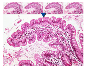
Brightfield, Phase Contrast, and DIC (Differential Interference Contrast) methods provide maximum options for your experiment.

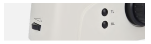
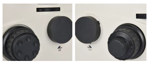
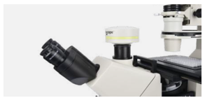
Convenient Lighting Control
The control buttons of the NIB900 Scientific Inverted Microscope are well-placed and easy to use. These buttons can simplify the workflow. For example, the transmission illumination switch and epi-fluorescent illumination shutter control button are placed on the right side of the machine.

Viewing Head with Bertrand Lens
The built-in Bertrand lens, moving in the optical path, can be used to observe the objective center and performs the same function as a centering telescope.

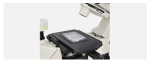
Tiltable Illumination Frame
The tiltable illumination frame provides a large working space for the user to change samples.
Removable Mechanical Stage
The high-performance three-layer mechanical stage is flexible and precise, equipped with a variety of stage mounting arms to accommodate different culture bottles and cell petri dishes.
Various Image Output Ports
An optical path output selection turntable is placed on the left side of the microscope. This facilitates the distribution of optical images to different ports and provides additional space for more optical image applications.
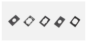
Load Stage
Equipped with a Terasaki holder, 96-well plates, Ø38mm, and Ø54mm petri dish holders, it meets various experimental needs.
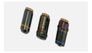
Camera Adapter
It provides 0.4x, 0.5x, and 1x C-mount options for users to choose from and is used to connect the camera with other image acquisition systems.
NIB910 STANDARD MODEL
Modular Design Provides a Variety of Flexible Imaging Methods
DIC Differential İnterference Contrast
DIC (Differential Interference Contrast) is a cost-effective optical technology that does not require expensive optics. It uses only a brightfield objective and two phase contrast adjustment sliders. For thicker samples, such as induced pluripotent stem cells, DIC provides a pseudo-three-dimensional, glare-free image. Halo effects, typically seen in traditional phase contrast observations, are minimized. Additionally, DIC can use glass culture dishes, making it a highly applicable observation technique.

NIB910-FL FLORESANS MODEL
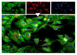
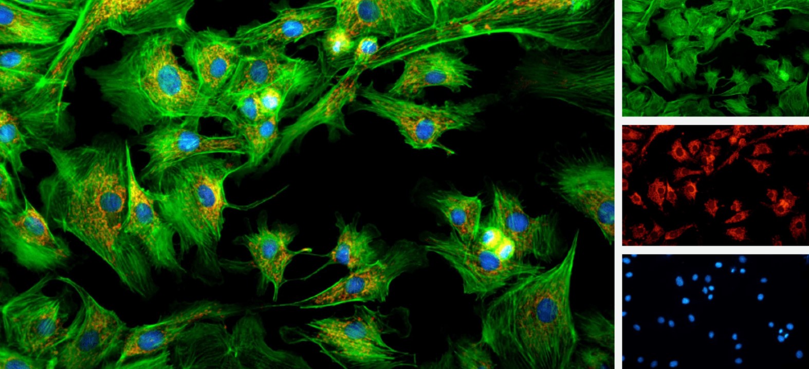
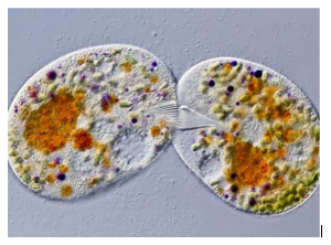
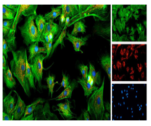
Provides You with Reliable, Clear, High-resolution Fluorescence Images
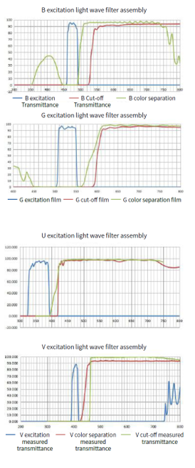
Uses the Latest Coating Technology
Using the latest advanced secondary corrugation elimination coating technology, the image becomes sharper, and the fluorescence transmission rate and detection efficiency are higher.
More Comfortable Fluorescence Observation
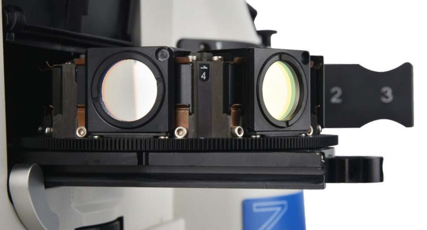
All fluorescent filter components are equipped with ultra-high-performance color filters. The fluorescence lighting support structure has a capacity for six color filter turrets, enabling the simultaneous imaging of differently stained specimens. High-sensitivity fluorescence achieves bright and high-contrast imaging results. Leading coating technologies reduce scattered light and spontaneous fluorescence, providing a higher signal-to-noise ratio.
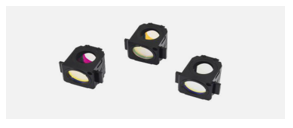
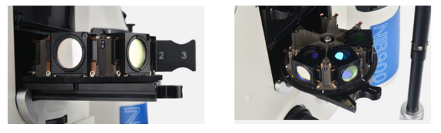
Fluorescence Filter Turret: Easier and More Flexible
The multi-functional six-station rotary structure can be easily removed from the main unit, allowing for the easy replacement of various fluorescence excitation modules.
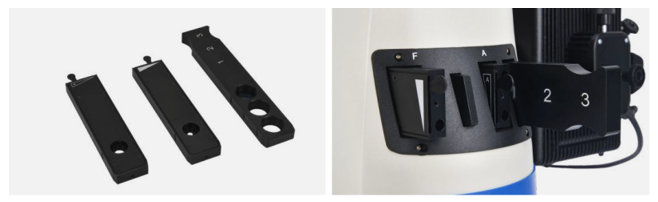
Simple and Fast Operation, Diaphragm Slider
In the epi-illumination optical path, three different light sliders—field diaphragms, aperture diaphragms, and filter inserts—demonstrate the versatility of the NIB900 in live cell research. When used with aperture diaphragms and fluorescence filters, the optimal fluorescence intensity can be adjusted according to the selected fluorescence module and objectives.
Measurement Function
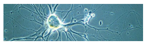
In cell and slice observations, measurement functions are required to determine cell size, cell gap, synaptic length, and other data. NOMIS Basic provides measurements such as distance, angle, rectangle, circle, ellipse, etc
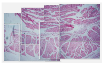
Quick Stitching
By acquiring and importing images in real time, NOMIS Basic can quickly stitch them together to create a large, high-resolution image.
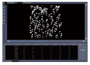
Cell Count
You can customize cell counting, automatically count cell shape information, and include data such as size, location, volume, perimeter, and brightness. All data and processed images can be saved as EXCEL tables.
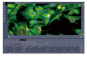
Fluorescence Image Synthesis
By acquiring or importing images from different fluorescence ports, the user can obtain an image after fluorescence synthesis. For each port image, fine adjustments can be made by adjusting the displacement in the X and Y directions.